How virtual hearts can save real hearts

Patricia Martínez Díaz 

4. May. 2021

In previous blogs, we talked about the theoretical basis for understanding how virtual hearts are made. We described the Hodking-Huxley mathematical models and how they helped us to improve our knowledge of the electrical activity of cells. In fact, those equations were the basis for obtaining the mathematical models to simulate the heart cells. In this post, I will explain how virtual hearts can be used to save hearts in the real life.

1. Hearts made out of numbers

In a very general way, you can now imagine that when we talk about virtual hearts, we are referring to the use of equations to model the functioning of the heart. This model will be then a simplification of the complexity of the cardiac organ and therefore, certain adaptations will have to be made to try to make it as close to reality as possible. One of the adjustments consists of incorporating patient information in order to have a personalized model. For example, we could use magnetic resonance images of the patient’s heart to generate the geometry of the organ, we could also incorporate information about the patient’s gender by modifying certain parameters in the equations. Additionally, the patient’s age could also be taken into account, since it has been observed that over the years the heart can develop fibrotic tissue. In general, the virtual heart should be as simple as possible, but at the same time not so simple that it is too far away from the real model and not so complex that it is too complicated to interpret it.

Figure 1. Building a personalized virtual heart form patient's data. Created in Biorender.com

The scientists’ vision consists precisely in creating a digital twin, which would be equivalent to a virtual heart of the patient that would allow us to generate a unique model adapted to his or her particular characteristics. Once with this personalized heart, the physician could then evaluate the best therapeutic option for that particular patient, for example to plan a surgery with the digital model or to determine whether certain drugs might be better compared to others. One of the areas where virtual hearts have been used is in cardiac electrophysiology, which studies electrical disturbances in the heart. Let me explain to you a little more in the following paragraphs.

2. Digital Twins to Treat Atrial Fibrillation

Atrial fibrillation (AF) is one of the most common heart rhythm disturbances in the population. In this disease, the upper parts of the heart (called the atria) beat at a very fast rate due to chaotic and irregular electrical activation. This prevents the lower chambers (ventricles) from filling with blood properly. Most of the time this disease goes unnoticed by patients as it is usually asymptomatic. The main problem is that during the rapid electrical activity, the blood can become stagnant and generate clots that travel either to the lungs or to the brain, endangering the patient’s life.

Figure 2. Personalized computer model of the atria

Treatments for atrial fibrillation consist mainly in the use of medications to control the heart rate or minimally invasive interventions where large tubes called catheters are inserted into the heart to eliminate the sites where the chaotic activity is generated. Despite being one of the most common rhythm disturbances, success rates for the treatment of AF remain low. This means that many patients continue having atrial fibrillation despite medical treatment. For this reason, computational models offer an alternative to personalize the treatment of atrial fibrillation and try to improve success rates.

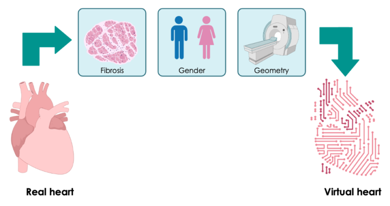
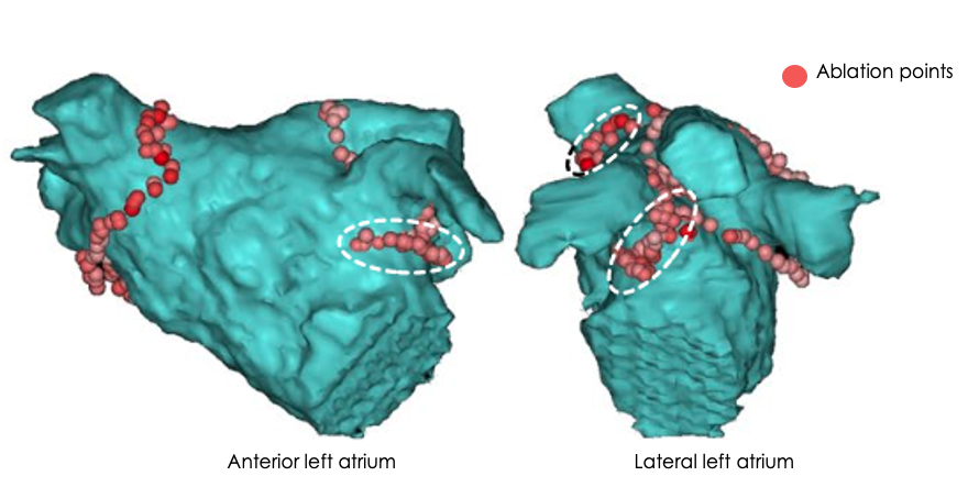
There are some studies [1,2] in which virtual hearts have already been used to plan procedures to treat atrial fibrillation. The main idea is to generate a digital heart capable of simulating the patient’s atrial fibrillation. Then with this model, the physician can test and ablate (i.e. burn) certain areas within the heart, just as in real life. After each test, it can be evaluated in the simulation if the arrhythmia has ceased, if not, a new ablation pattern can be generated and retested if the heart continues to beat chaotically or not. Once the optimal sites that will potentially eliminate the arrhythmia have been identified, a map is generated which can be superimposed on top of the image of the virtual heart and then be projected on the electrophysiology lab screens during the actual ablation.

Figure 3. Location of ablation points on a biatrial model as shown in Boyle et al. 2019

I hope that with this blog you have a better idea of what virtual hearts are and how they can help us plan treatment for atrial fibrillation. Thanks for reading!

3. References

  1. Boyle, P. M., Zghaib, T., Zahid, S., Ali, R. L., Deng, D., Franceschi, W. H., . . . Trayanova, N. A. (2019). Computationally guided personalized targeted ablation of persistent atrial fibrillation. Nature Biomedical Engineering, 3(11), 870-879. doi:10.1038/s41551-019-0437-9
  2. Shim, J., Hwang, M., Song, J., Lim, B., Kim, T., Joung, B., . . . Pak, H. (2017). Virtual in-silico modeling guided catheter ablation predicts effective linear ablation lesion set for longstanding persistent atrial fibrillation: Multicenter prospective randomized study. Frontiers in Physiology, 8. doi:10.3389/fphys.2017.00792


Follow us in our social media accounts to stay tuned, and contact to personalizeAF@itaca.upv.es if you want to know more about our project!