Computer models of the atria: first contact and challenges

Get to know one of the models of the atria developed by the PersonalizeAF members in Maastricht

Atrial fibrillation is a very complex and dynamic arrhythmia. Understanding the conditions for its maintenance and finding the most effective treatment strategies are challenging tasks. One very important tool to help clinicians and researchers achieve these goals is the measurement of electrical signals directly in the atria, which can help elucidate the arrhythmic mechanisms.  However, experimental data may be insufficient to gain in-depth knowledge of the arrhythmia, as the procedures are often limited by the available technology or patient safety. One possible way to complement the knowledge obtained from experiments is to use computational modeling, which is one of the pillars of my work here in PersonalizeAF and the topic of this month’s post.

Computers have been used to simulate atrial fibrillation since the 60s, helping establish some important developments in the field, such as the hypothesis (for a long time unchallenged) that multiple wavelets are the mechanism behind the arrhythmia. Many modeling strategies have been developed, incorporating progressively more complex anatomy and electrical dynamics (check this review for more info on that).

Currently, some of the most complete models of atrial fibrillation are developed by members of PersonalizeAF, including Karlsruhe Institute of Technology, Université de Bordeaux, and Maastricht University. The focus of this article is the model developed in part by researchers in Maastricht and at the Univeristà della Svizzera Italiana, with which I have been working since my start in this project.

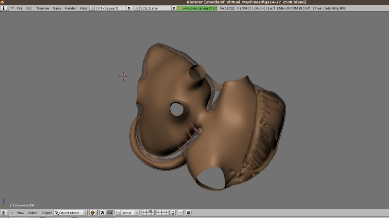
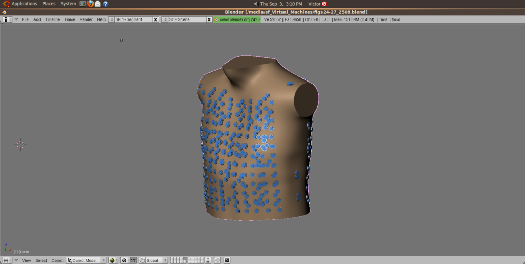
This model, which is very nicely detailed in this paper, includes important anatomical features that help achieve realistic electrophysiological simulations. It is based on magnetic resonance imaging of a patient’s atria, complemented by details added manually in Blender, such as bundle networks. The cells are oriented following anatomical studies, accounting for the natural anisotropy along with the atrial muscle. The model is capable of generating signals not only in the atria but also anywhere on the torso, enabling the researchers to explore non-invasive signals such as electrocardiograms, body surface potential mapping, and transesophageal ECGs.

The Blender interface with the multi-layer atrial model developed at Maastricht University and Univeristà della Svizzera Italiana

Instead of a simpler approach of simulating a single layer of cells in a 3D shape, this model has multiple layers between the epicardium and endocardium, accounting for a much more detailed, truly tridimensional electrical propagation. This is a unique feature of this model, which enables the investigation of arrhythmic mechanisms that involve both layers of the cardiac muscle, such as intramural conduction.

The conduction of each cell is, similarly to other cardiac models, based on mathematical descriptions of the ionic currents that are involved in the propagation of electrical currents in the heart. This allows the specification of the electrophysiological characteristics of individual portions of the atria, which may be more or less conductive. This is important to allow the model to reproduce the heterogeneity observed in atrial fibrillation patients, either related to the presence of fibrotic tissue or differences in electrical properties.

The current version of the model allows the investigation of possible factors that favor the appearance of atrial fibrillation drivers, as well as exploring the effectiveness of ablation and drug therapies. One of my goals in PersonalizeAF is to contribute to the massive amount of work that has been made so far in this model.

Our model can simulate the electric activity of the heart also on the torso surface and esophagus, reproducing non-invasive measurements

We hope to make this model more capable of representing the individual characteristics of each patient so that it can then be used to explore the treatment possibilities in an environment without risks. This involves incorporating more spatial variability in electrophysiological and structural properties, aiming to achieve more realistic conduction patterns. We also want to use the models to explore the relationship between regions driving the arrhythmia and the many signal processing strategies that can be applied to both invasive and non-invasive data.

So far, I have been working to wrap my head around such a complex topic, which is being a very interesting journey. I started learning Blender, which is a fascinating and very complete open-source software, and I worked to obtain simulated electrogram signals from the model, mimicking a real invasive measurement, so I can start running some analyses on them.

I hope I can get some interesting results to share as soon as possible! Until then, be sure to follow PersonalizeAF on Twitter and LinkedIn to remain updated about the project, including the developments of my work and that of the other ESRs.


See you soon!

Victor