Using Computer Models to Study the Heart

Patricia Martínez Díaz 

1. December.2020

Welcome to my blog!

Do you imagine a world where doctors have access to a digital version of their patients’ hearts and can use it to plan procedures before entering the operating room? This might sound like a dream, but it is becoming true. In this blog I will talk about the use of computer models in the field of cardiac electrophysiology and I will also share with you my journey as a PhD student at the Karlsruhe Institute of Technology. Welcome to the world of computational cardiology. Happy reading!

1. What is a model?

To understand how to build up a digital twin of the heart and how it can be used to plan patient´s therapy, it is important to understand how scientists came up with the idea of modeling the heart. First of all, I would like to give you some background information. In general terms, a model is a representation of a complex system. A model can be either a schematic diagram of the phenomenon, as the planes of a new house, it can also be a physical model such as a miniature car, or even a mathematical equation. In other words, if a tool is used to help us understand the functionality of the object under study, then it can be called a model.

Figure 1. Simplified model of the systemic circulation described by Guyton (1). This schema is used to explain how the changes in arterial and venous resistance affect the quantity of blood that the heart pumps.

2.The Squid that helped us understand the cell

More than 80 years ago, in 1939 Hodgkin and Huxley (2) proposed the first mathematical model describing the voltage changes in the cell membrane caused by modifications of the ionic channels (small “doors” that allow or block the flow of ions into and out of the cell). They derived a series of mathematical equations from experimental data obtained from the nerve fibers of a squid. The voltage fluctuation is called the action potential.

A good way to understand the action potential is to imagine the cell in two different states: at rest and at the excitation phase. When the cell is resting it has a negative charge with respect to the surrounding space. However, if the cell is properly stimulated, it changes its polarity, meaning that it becomes positively charged. In Figure 2 you can see a representation of the experiment performed by Hodgkin and Huxley and the original action potential recording found in the original paper. By the way, in 1963 together with John Carew Eccles, they were awarded the Nobel Prize in Medicine (3).

Figure 2. A)Experiment performed by Hodgkin and Huxley to obtain the action potential of the squid axon. Adapted from (4) and Biorender.com
Figure 2B. Experiment performed by Hodgkin and Huxley to obtain the action potential of the squid axon. Adapted from (4) and Biorender.com

3. The first model of a cardiac cell

In general terms, both neurons and muscle cells can be classified as excitable cells. The heart has its own muscular cells, also known as cardiomyocytes. They need electricity to be able to contract and pump the blood through the body. These electrical impulses come from the specialized cells known as the electrical conduction system. These cells have the ability to start and propagate the stimuli to the rest of the cardiac tissue.


In 1960 Denis Noble (5) obtained a model to characterize the action potential of a cardiac electric cell. As seen in Figure 3, the shape of the wave is different from the one obtained by Hodgkin and Huxley. This is due to the fact that for cardiomyocytes, calcium needs to enter from the extracellular space to allow their contraction, this is represented by the long flat plateau in the middle of the signal. Since then several models have been developed describing the electrical function of the cardiac cells.

Figure 3. Original cardiac action potential computed from the model developed by Denis Noble. Adapted from (5)

4. Scaling: From single cell to modeling the whole heart

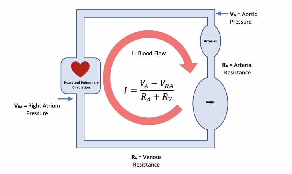
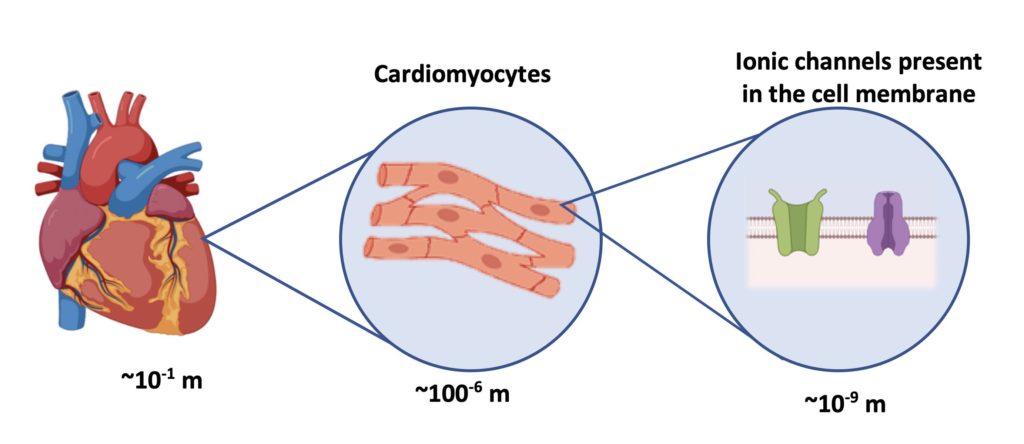
In order to reach our goal of simulating the whole heart, there are more points to think about. First, consider that the human heart is made up of billions of cells (6). Consider also that not all of the cells belong to the same group, this means that the heart has different types of cells, including electrical and muscular, which behave differently and therefore have distinctive types of action potentials. The challenge is to scale the model to be able to represent the entire organ as shown in the figure below. As you can imagine this is not an easy task, for this reason we need computers to help us solve the equations, but we will discuss this later in the next post.

As we get closer to answer our question on how to model the heart, we will learn more about its function and also about modelling and simulation tools. Thanks for reading.

Keep safe and see you soon.

Figure 4. Spatial scaling and the challenge of whole heart modelling. Biorender.com

5. References

  1. Guyton AC, Abernathy B, Langston JB, Kaufmann BN, Fairchild HM. Relative importance of venous and arterial resistances in controlling venous return and cardiac output. Am J Physiol-Leg Content. 1959 May 1;196(5):1008–14.
  2. Hodgkin AL, Huxley AF. Action Potentials Recorded from Inside a Nerve Fibre. Nature. 1939 Oct 1;144(3651):710–1.
  3. The Nobel Prize in Physiology or Medicine 1963 [Internet]. NobelPrize.org. [cited 2020 Nov 26]. Available from: https://www.nobelprize.org/prizes/medicine/1963/summary/
  4. Petkov GV. Chapter 16 – Ion Channels. In: Hacker M, Messer W, Bachmann K, editors. Pharmacology [Internet]. San Diego: Academic Press; 2009 [cited 2020 Nov 26]. p. 387–427. Available from: http://www.sciencedirect.com/science/article/pii/B9780123695215000166
  5. Noble D. Cardiac action and pacemaker potentials based on the Hodgkin-Huxley equations. Nature. 1960 Nov 5;188:495–7.
  6. Tirziu D, Giordano FJ, Simons M. Cell Communications in the Heart. Circulation. 2010 Aug 31;122(9):928–37.