A Tour de Force for Atrial Cells

Introducing you to cell isolation, calcium handling and the magic of life.

These last two months have been crazy busy with experiments. The project I am working on right now aims at investigating local calcium releases, events called “calcium sparks”, after fast electrical stimulation of atrial cells to mimic atrial fibrillation.

If you are interested in how this is done, allow me to guide you through the procedure. The first step is, of course, to obtain good atrial cardiomyocytes (aCM).

Just for you to have an idea of the anatomy of the atrium, it can be divided in two parts: free wall and appendage (Figure 1). The former, is thicker and “denser” compared to the latter, and it is the portion attached to the ventricle. The appendage instead, is the “flap” of the atrium, free to move and with a pocket-like structure; it has lots of trabeculae, in order to be able to resist the blood load despite the thinner structure.

Figure 1 - Left atrium. This side of the atrium is normally not visible because facing the ventricle. The access to the appendage (white arrow) is how ventricle and atrium communicate. On the right, you can see the difference between the trabecular structure of the appendage (bottom) and the denser structure of the free wall (top).

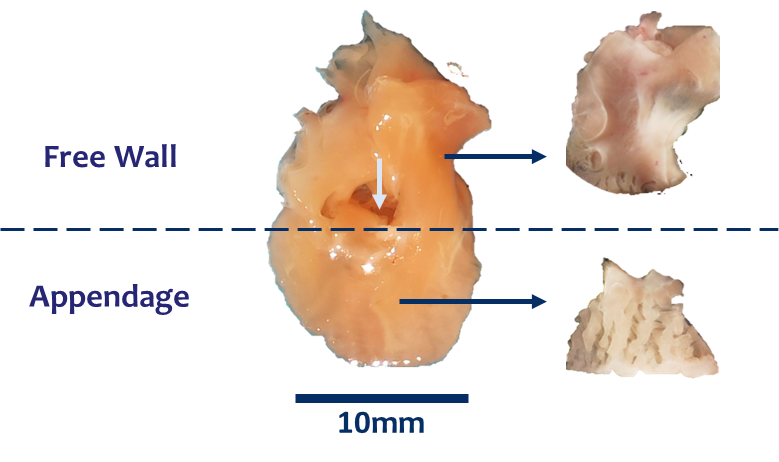
Now, given the tissue, I need to obtain single cells. However, you may have already thought that with one atrium there are not many conditions I can simultaneously test. This is the reason why slicing the tissue comes in so handy; from one atrium I can test multiple conditions.

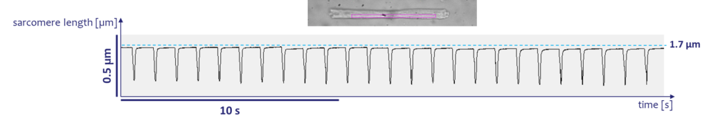
After obtaining slices, I can reach one of the key steps of the story: cell isolation! This technique consists in several steps of chemical digestion of the tissue. Once I have obtained singles cells and restored physiological conditions, it is time to assess their health status. As you can see from the video below, when mimicking the action potential via electrical stimulation, atrial cells respond by contracting synchronously (Video 1). A further proof of cell responsiveness is obtained by recording sarcomere shortening traces, where you can clearly see the amplitude of the contraction (Figure 2).

Video 1 – Sarcomere shortenting. Isolated atrial cardiomyocytes respond to electrical stimulation by contracting synchronously.

Figure 2 - Sarcomere shortening. The trace shows the amplitude of contraction of one atrial cardiomyoycte during electrical stimulation.

If cells have a good sarcomere length (it should be above 1.7 μm), they are good candidates for calcium imaging experiments. Calcium handling is what allows cell contraction and therefore the pumping activity of the heart. If a cell is healthy, each contraction is associated with a synchronous calcium release throughout the cell, as it was suddenly switched on (Video 2). However, we are interested in atrial fibrillation, aren’t we? This is a pathological state, which from the calcium handling perspective translates into calcium waves and sparks. This leads to an arrhythmic behaviour, where occasional localized calcium releases (sparks), seem to lead to calcium waves which travel from one side of the cell to the other. Stimulating cells at a frequency that matches atrial fibrillation, allows inducing this pathological behaviour.

Video 2 – Calcium signal. When an atrial cardiomyocyte is paced, it contracts as a consequence of synchronous calcium release throughout the whole cell.

 Frangar non flectar is a roman motto which literally means I may break, but I won’t bend. It is however commonly used to express the energy and strength pushing someone not to break in front of adversities. I see these cells as the exemplification of this way of saying; after “torturing” them for hours, they still contract synchronously and regularly! What would you do if you were sliced, chemically digested, moved from one dish to the other and electrically stimulated? I would definitely be very stressed and not willing to do absolutely anything of what I would have otherwise done. I know, this comparison does not nail the point, but it does give an idea of how powerful life is and how these little tiny cells we are made of can endure such adverse conditions. Atrial cardiomyocytes may look a bit crooked after isolation sometimes, but they proudly show off that they are still there, not willing to give up!

I have to go and check how my cells are coping now, see you in the next post!

Teresa

Do not forget to regularly check #PersonalizeAF website, LinkedIn and
Twitter to have monthly updates about the latest achievements
of the network in the field of #AFib research!