1.Teaching tools
An early application of 3D printing cardiac model was to build models for anatomy instruction or presentation. Similar plastic heart model known to most healthcare authorities, the 3D printing model can speedily reproduce multipart anatomical structures, but it is said to illustrate the anatomical pathology of the patient. There is also an added value. These models can be used as instructions to guide healthcare professionals about the connection between ordinary and irregular structures, as well as help the general public to better understand some structural heart diseases.
2.Functional flow models
The combination of high-resolution spatial tomography, CAD software, and multi-material 3D printing technology makes it easy to create 3D models of aortic regurgitation for patients. Aortic valve dysfunction is a recently repeated series of 3D printing associated with a flow model. Severe aortic stenosis (AS) indicates that the valve structure is relatively stable, so CT data sets show the anatomy of the aortic root, including the valve opening region and local calcification. You can get patient-specific details of the anatomical structure.
3.Procedural planning
Congenital cardiovascular disease is usually accompanying with intricate and exclusive geometric shapes that are tricky to fully understand from a 2D CT, MRI, or echocardiogram. Therefore, 3D printing modeling can be an important role in supporting a more inclusive appreciation and efficient assessment of various congenital heart disorders.
The latest applications of 3D-printing congenital heart models include pre-operative planning and interventional simulation, using aseptic models intraoperatively to improve the structural orientation, and functional hemodynamics of the patient. Include evaluation and testing of new procedural methods. Complex congenital cardiac anatomical structures such as double right ventricular defect, atrial septal defect (ASD), ventricular septal defect (VSD) and 3D printing of surgical planning were reconstructed.
Application of 3D printing in Atrial fibrillation:
Atrial fibrillation is the most common type of arrhythmia and the main cause of stroke. Given that more than 90% of left atrial fibrillation (LAA) thrombosis occurs in patients with non-valvular atrial fibrillation, percutaneous LAA occlusion can effectively prevent patients with stroke-induced atrial fibrillation, especially the heart. Suitable for high-risk patients with thromboembolic stroke, but oral administration is prohibited. Anticoagulant. However, the structure of the LAA is so complex that adequate information about it is essential for successful LAA closure.
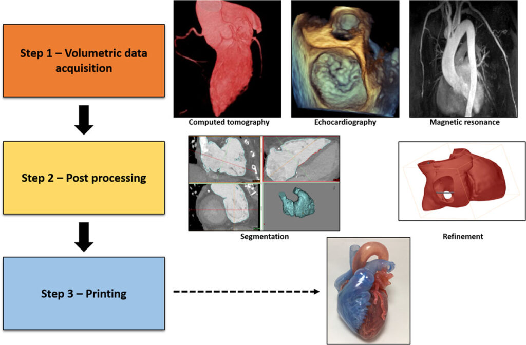
3D printing technology has made great strides in the past decade and has been used in a variety of biomedical applications. Using a 3D printed heart model eliminates this possibility and removes the spatial aspect of the imagination. This is invaluable for patients with complex anatomical structures. The data obtained from CT were reported to have succeeded in generating a 3D model of the LAA. However, the use of real-time 3D TEE data for LAA 3D printing has not been studied. Compared to cardiac MRI and CT, TEE has several advantages. They are easy to carry, easy to obtain, have high time accuracy, require no radiation, and can be operated without sedation or conscious anesthesia (if requested to do so). Currently, TEE is regularly performed for LAA obstruction. Therefore, the use of 3D-printed LAA echocardiographic data provides a new level of clinical accessibility. Below is the figure which shows typical workflow of 3D printing medical data.