How does your heart beat?

We all know our heart beats, but how? What affects this beating?

Since the beginning of our lives, we have been told about our heart beating. We grow up knowing we have some kind of pump inside our chest that beats and beats and beats and it never stops beating (luckily for us!). Sometimes we put our hand on the chest to feel this pump, and sometimes we feel it slowlier or faster, depending on the situation. But does it beat homogeneously? Is there any kind of structure that makes your heart beat in one way or another? Does your heart beat the same way on each part of its body?

Maybe I asked too many questions, but I’ll try to go over them in this post. First of all, let’s start with the basics. The heart does not beat homogeneously in all of its body. We all know there are two systems in the heart: the left and the right one. In primary school we were taught that the right part of the heart is involved in filling the blood with oxygen and the left part of the heart is the one involved in distributing the blood to the rest of the body. To do so, both sides of the heart have an atrium, which receives the blood, and a ventricle, which sends out the blood. I am sure you all remember all of this from primary and high school. Let’s go a bit deeper into this.

When the heart is healthy, its muscular body is made out of cardiomyocytes, cells that have a beating function. However, when we grow older or when we have some kind of disease, the muscular body gets infiltrated with other types of cells: fibroblasts, which are scarry cells. These are the cells that, when we have some kind of injury end up recovering the tissue. As you can imagine from the scars you have seen in your skin, the tissue made out of fibroblasts is different than the original one. The same can happen in your heart. In the picture on the right you have a visual example of this difference in tissue, of course seen by dying scarry tissue.

Difference in cardiac structure due to fibrosis caused by fibroblasts [1]

As you may be thinking, if the muscular tissue is made of cells other than cardyomyocytes, the beating function might be affected. This can be seen in the figures below. Whereas on the figure of the right a smooth activation throughout the entire surface of right and left atria is observed, on the left there is the wavefront propagation in the left atrium of a patient with atypical atrial flutter, an arrhythmia that affects the propagation of the electrical impulse and makes it less homogeneous and cyclical. We can see how the conduction is slowed on the posterior wall area, close to the left inferior pulmonary vein, where the wavefront needs more time to reach the lateral wall.

Wavefront propagation on an endocardial activation map of the left atrium. Patient with atypical atrial flutter
Wavefront propagation of both atria on a healthy heart [2]

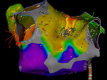
When we see something like this, this area is labeled as slow conduction area. If we analyse the conduction velocities of this area, we will find how the values of this area are lower than those of the atrial roof or the central area of the posterior wall. 

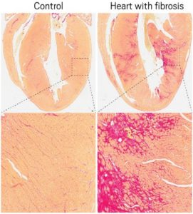
This beautiful and coloured map that shows this slowing down of the propagation of the wavefront, however, is obtained during a surgical procedure. It would be ideal to obtain information about slow conduction areas without the need to perform invasive procedures, even minimally invasive procedures, as in this case. And this is what we are trying to study. I am trying to correlate the information obtained with this invasive mapping tool to two other non-invasive techniques, which are MRI and ECGi. In the case of ECGi, I would be comparing it in terms of conduction velocity as well, or perhaps with dominant frequency maps, but in the case of MRI, since it gives anatomical and not functional information, I would be comparing it to fibrotic areas, which is something that has been studied and has been seen to have a negative correlation [3]. If we find similar results by using other methods to calculate conduction velocities, and we can correlate these three techniques information, our results may facilitate the targeting of substrate for reentrant arrhythmias by using non-invasive techniques. We shall see in the future!

For the moment, I hope you got clear from this post that the heart does not beat homogeneously in all of its body, that its beating is affected by scarry tissue, which affects differently every part of the heart, and that this affects the homogeneity of the wavefront propagation. In other posts I will try to explain methods to calculate conduction velocity by using activation information from the electroanatomical mapping, and how am I trying to correlate this information to MRI information and ECGi information. It is a Friday afternoon when I am writing this and it is sunny, so allow me to take a break from research to go for a walk and enjoy some sun. See you in other posts!

Eric


If you want to join me and the rest of the network in our journey, don’t forget to follow us in our social media using the hashtag #PersonalizeAF!